Harry is a 61-year-old man who comes in for a mild URTI and is incidentally noted to have a blood pressure of 178/120 mm Hg on examination for this. You have no further history on this gent as he is new to the practice. You perform fundoscopy in context of cardiovascular examination after measuring this BP with image as below:

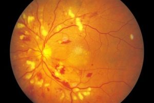
Describe the Features seen on this fundoscopic picture (5)

  1. Generalized arteriolar narrowing
  2. Focal arteriolar narrowing
  3. Arteriolar wall opacification,
  4. Arteriovenous nipping
  5. Flame-shaped hemorrhages
  6. Blot-shaped hemorrhages
  7. Cotton-wool spots
  8. Hard exudates
  9. Microaneurysms
  10. Optic disc swelling (papilledema)


What are your differentials for this presentation? (2)

  1. Hypertensive retinopathy
  2. Diabetic retinopathy
  3. HIV retinopathy
  4. Optic neuritis

References

  1. patient.info
  2. Murtaghs
  3. Colour Atlas of Family Medicine
  4. Dynamed