
Sally is a 25 year old Caucasian woman who presents with loss of vision over a few days in one eye, associated with eye discomfort on movement of the eye, with changes in vision and difficulty focussing directly onto objects, reduced colour perception, flashes in the eye on eye movements. Fundoscopy is only remarkable for an optic disk that looks as shown.
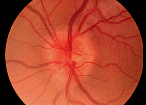
What is the most likely diagnosis given the history described? (1)
What are the key features of history in the condition described? (5)
- Visual impairment
- Pain around the eye
- Pain worse on eye movement
- Dyschromatopsia
- Scotoma
- Light flashes with eye movement
NB – in the RACGP KFP it is VERY UNLIKELY you will receive marks for having answers that are already given in the stem.
What are the key features of examination in the condition presented? (5)
- VA reduced
- Scotoma
- Relative Afferent pupil defect on affected side.
- Optic disc swollen
- Optic nerve atrophy
- Disc pallor – occurs 4-6 weeks later
- Normal macula and peripheral retina
What are the key investigations in this condition? (5)
What are the key features of management in this condition? (1)
What are the complications of this condition? (4)
- May be first presentation of MS
- Optic nerve atrophy
- Optic neuritis in other eye
- Poor visual recovery
What is the most common cause/association of ADON? (1)
What are the most common causes of this fundoscopic finding in one eye? (3)
- Demyelinating optic neuritis
- Non-arteritic anterior ischaemic optic neuropathy
- Retinal vein occlusion
- Diabetic papillopathy
BONUS QUESTIONS
What are the most common causes of bilateral presentations of this fundoscopic finding? (3)
What are some conditions of the optic nerve that could cause the fundoscopic findings seen? (5)
- Optic neuritis
- Arteritic ischaemic optic neuropathy
- Giant cell arteritis
- Non-arteritic anterior ischaemic optic neuropathy
- Toxic optic neuropathy
- Methanol poisoning
- Compressive optic neuropathy
- Thyroid eye disease
- Congenitally anomalous optic discs
- Infiltration of the disc by sarcoid
- Infiltration of the disc by glioma
- Infiltration of the disc by lymphoma
What are some intracranial conditions that could cause the fundoscopic findings seen? (3)
- Brain Tumour
- Cerebral trauma
- Intracerebral haemorrhage
- Subdural haemorrhage
- Cerebral inflammation/infection
- Cerebral abscess
- Idiopathic intracranial hypertension
- Chiari malformation
- Acute mountain sickness
- High-altitude cerebral oedema
- Lyme disease
What are some medication classes that could cause the fundoscopic findings seen? (4)
What are some vascular conditions that could cause the fundoscopic findings seen? (4)
What are some globe conditions that could cause the fundoscopic findings seen? (5)
References
- patient.info
- Murtaghs
- Colour Atlas of Family Medicine
- Dynamed


