
Harry is a 70 year old man presented with acute persistent painless unlilateral loss of vision, starting like a curtain descending. He can barely see his hand in his right eye. He has a history of hypertension, diabetes, hyperlipidemia and AF.
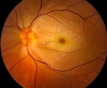
What are the features of fundoscopy you see in the above picture?
- Cherry Red spot on fovea
- Pale retina
What are the differentials of the above presentation?
- retinal artery occlusion
- retinal detachment
- Amaurosis Fugax
- retinal vein occlusion
- Stroke
- Vitreous Haemorrhage
What are the key features of history that would allow you to differentiate between causes of Harry’s presentation?
- History of diabetes
- History of recent trauma
- Cardiovascular history
- neurological symptoms
- History of AF
You are considering a diagnosis of Central Retinal Artery Occlusion. What are the key history feature of retinal artery occlusion?
- Sudden loss of vision like curtain descending
- Painless
- Unilateral
- Sudden
- Central vision lost
- Not improved 1mm pinhole
- No light perception
What are the Causes and associations of Retinal Artery Occlusion?
- Cardiovascular risk factors
- HTN
- Diabetes
- Hyperlipidaemia
- AF
- Atherosclerosis
- SLE in younger patients
- Wegeners Granulomatosis
- Behcets Disease
- GCA
- Hypercoaguable state
What are the key fetaures of examination in central retinal artery occlusion?
- VA not improved with 1mm pinhole
- VA markedly reduced
- retinal arteries emboli
- Red reflex abnormal and asymmetrical
- Cherry red spot on macula
- Relative afferent pupillary defect
- Attenuated arterioles
- Pale retina due to ischemia
What is the source of the emboli in central retinal artery occlusion?
- Carotids
- Heart Valves
What is the immediate managament of this condition?
- Urgent referral for treatment within 4 hours
What are the immediate steps in Managament of central retinal artery occlusion?
- Referral to ophthalmologist
- High flow inhaled 100% O2
- IV corticosteroids
- Massage globe digitally though closed eye closed within 30 min
- Anterior Chamber Paracentesis by eye surgeon
- Hyperbaric oxygen within 12 hours
- IV Acetazolamide 500mg to reduce IOP
- Timolol 0.5% topical drops BD to reduce IOP
SoundCloud Lecture
[wpforms id=”7496″ title=”false” description=”false”]


